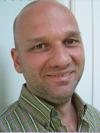
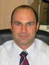
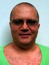
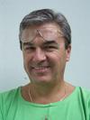
Matild Mayer works at Ars Medica Laser Clinic in Budapest, Hungary, a ISO-accredited center.
Data provided by the hospitalLast updated 18 Nov 2009
Profile under review. Education, experience, memberships and awards are not yet published for this doctor. The hospital's team can answer questions about the doctor directly.
Ask the hospital about this doctor
Send your reports and questions to Ars Medica Laser Clinic. The hospital replies through Health-Tourism.com.
Free consultation • No obligation • Direct communication with the hospital
Root-end resection is performed on outpatient basis, under local anesthesia (2% Lidocaine injection). With incisions around the tooth we create a flap, which we reflect, then drill a small hole into the bone around the apex with a bur. The opening is as large, as the infected area's diameter. Next, we remove 1/3 of the apex with a bur, and then perform an apical currettage. The next step is retrograde sealing of the obturation with cement. We smooth the sharp edges with a bur, return the flap to its original position, and close it with sutures.
On the day of the surgery, the patient signs the consent form, the necessary lab tests are done and the fee for the procedure is paid. Immediately before surgery the patient is premedicated with an injection containing analgesics and sedatives. The surgery may be done with local, spinal or under general anesthesia. local anesthesia means infiltration of the surgical field with 1% Lidocaine. The 4-5 cm incision can be made in the crease below the breast, in the armpit or across the areola. In the pocket that is created for the implant we frequently place drain tubes to allow the serous secretions to escape; these are removed the next day. The implant is inserted between the breast tissue and the pectoral muscle. Then the wound is closed in layers, skin closure is always done with cosmetic sutures. At the end of the procedure pressure dressing is applied to the chest.
The procedure involves correction of the position, shape and size of the breasts. If size is the main problem we remove the necessary skin and breast tissue; however if the size of the breasts is different, depending on the patients constitution and other factors, reduction of the larger breast or augmentation of the smaller with an implant may be performed. Correction of shape and position of the breasts is accomplished by removal of the excess lax skin and securing the breasts in their physiological position. Following careful hemostasis the wound is closed with cosmetic sutures. Frequently suction tubes are placed into the surgical area to drain serous secretions. At the end of the procedure dressing is applied to the chest.
We highly recommend eating before surgery, because after the injection of the anesthetic, eating is not allowed for 3-4 hours, or until the anesthetic wears off. We perform the procedure in local anesthesia, which implies infiltration of the surgical area with 2% Lidocaine. The dentist reflects the full thickness flap from the bone, and by using a special bur, establishes the depth and alignes the axis of the implant recipient site. Into this recipient site, the dentist screws in a titanium-based implant, which can be viewed as the root of the new tooth. We close the surgical wound with sutures. The screw placed in the bone will take 4-6 months to heal. During this time, the space between the titanium screw and the surrounding bone undergoes ossification, embedding the implant into the bone. The screw cannot be loaded during the healing phase. That's why a provisional bridge or denture can be made during this time. The second operation takes place when the screw is firmly embedded in the jawbone. Next, we expose the screw from underneath the gum tissue and place a small head on it, which elevates it above the gum. We call this a healing cap. The gum will heal for 2-3 days, then the last phase of treatment takes place, which does not require surgery. During this phase we attach an abutment, which serves as the foundation for the future bridge. Following this and the impression, the dental technician will construct the appropriate restoration in multiple phases.
Fabrication of bridges for combination works is explained in the crowns and bridges section. All types of dentures are made in a similar way. During the first visit, we take preliminary impressions of both the maxillary and mandibular arches, which allows the technician to make a custom tray. This helps us attain a more precise impression and fabrication of a more stable denture. During this visit, we also measure the central occlusion. This is important, because we are able to fill the void left by the missing teeth with a wax rim, which supports both the face and the lip, eliminating the wrinkles associated with a caved in mouth. During the next phase, we try in the teeth in wax. At this time we are still able to modify the size, shape and position of the teeth according to the patients wishes. Adjustment of the teeth in wax, requires another try in. Finally, when all teeth are esthetically correct, the dental technician completes the final denture. The whole process takes about two weeks form the first impression, or three weeks in case of combination work.
The technician guarantees the denture for a year. The interim denture is not included in the warranty due to its temporary nature. The warranty is only valid, if the patient appears at the half-year follow up examinations.
The procedure may be performed under general anesthesia, or using local anesthesia (infiltration of the surgical field with 1% Lidocaine). During the procedure, we obtain tissue scrapings from two different sites: the uterine cervix and the uterus. The samples are carefully labeled for site of origin and sent to pathology. The procedure also provides information on the size and possible gross abnormalities of the uterus or the uterine wall (myoma).
On the day of the surgery, the patient signs the consent form, then the necessary lab tests are done and the fee for the procedure is paid. Immediately before surgery the patient is premedicated with an injection containing analgesics and sedatives. The surgery may be done with local or under general anesthesia. Local anesthesia means infiltration of the surgical field with 1% Lidocaine. After an incision is made at the back of the ear the cartilage is reoriented into the desired position. If necessary an excision of appropriate size is made from the cartilage, which is then secured in the new position. Following hemostasis the skin is closed with cosmetic sutures. After surgery a pressure dressing is applied.
The procedure is usually performed in local anesthesia but it can of course be done under general anesthesia as well. During plastic surgery of the upper eyelid an incision is made in the upper part of the eyelid, hidden in a natural crease, followed by excision of the excess skin marked for removal previously. During plastic surgery of the lower eyelid the incision is made 1-2 mm below the lashes parallel to the line of the eyelid. Reduction of the skin is usually moderate and the accumulated fat has to be removed in most cases. If necessary we also perform fixation of the supporting tissues. Wound closure is done with hidden cosmetic suture. During plastic surgery of the lower eyelid in certain cases removal of fat is possible through the conjunctival aspect of the eyelid leaving no visible scar at all. Plastic surgery of the upper and lower eyelids can be performed during separate sessions or simultaneously. Frequently plastic surgery of eyelids is done simultaneously with surgery for corrections of wrinkles on the face. At the end of the procedure we secure the sutures with bandaids.
The procedure is usually done with local or under general anesthesia. Local anesthesia means infiltration of the surgical field with 1% Lidocaine. The procedure requires lengthy, careful preparation therefore patients need to be premedicated with a sedative-analgesic injection prior to procedures with local anesthesia. The incision begins above the hairline at the temple, continues in front of the ear curving back behind the earlobe to the nape where it again continues in the hairy scalp, mostly in areas covered with hair. Therefore after wound healing the scars are covered with hair and are not visible. During the procedure the facial skin is tightened after mobilization of the subcutaneous connective tissue and the excess flabby skin is trimmed. The wounds are closed with cosmetic sutures and a pressure headband is applied.
Intervention can be carried out with narcozitation (in childhood anyway) or with local anaesthetic. From these two opportunities patients have to choose based on the received information, discussion with doctors. Surgeon removes the extended mucosae with the help of a special optical equipment so-called endoscope through nostril beside eye check.
After surgery - carried out either in local or general anaesthesia - for monitoring reason one night must be spent at the clinic.
On the day of the surgery, the patient signs the consent form, then the necessary lab tests are done and the surgery fee is paid. Immediately before the operation, the patient is premedicated with an injection containing analgesics and sedatives. The operation can be performed under local, spinal or general anesthesia. The majority of our surgeries are performed with local anesthesia, which means infiltration of the surgical field with 1% Lidocaine. Next the hemorrhoidal nodes located in the typical directions are removed with laser. The procedure leaves three non-bleeding wounds, covering about 4X1 cm. The laser practically seals the incision, eliminating the need for sutures, thus the wounds remain "open". This allows the post-surgery edema to drain unhindered thus prevents development of tension, which greatly reduces post-surgery pain, allowing the patient to be discharged on the day of surgery.
The duration of surgery is 15-25 minutes.
The operation is generally carried out by local anaesthesia, rarely in genaral anesthesia. Local anaesthesia means injection of the operative area with 1% Lidocaine solution. The intervention is diuturnal therefore before local anaesthesia a so-called preparing injection consisting sedative and pain killer is given. During the operation the enraged labia minor - due to the previously defined lines- are reduced ibn size and following the careful haemostasis the wound is closed by plastic stitches.
Prior to surgery the surgeon exactly marks the sites for liposuction. This is followed by local anesthesia or general anesthesia. This is followed by injections of a medicated solution into the previously marked areas that serves to dissolve and loosen up the fat. The surgical intervention starts from one or more small, approximately 4-5 mm incisions on the skin. Through these small openings suction cannula is introduced under the skin and with the help of vacuum the excess fat is suctioned off. The procedure is repeated in all the marked areas, then the entry openings are closed with fine sutures. At the end of the surgery pressure dressing and a snug compression garment is applied.
Regardless of the reason for root canal treatment, the procedure is always similar. After the local anesthesia of the teeth (with a 2% Lidocaine injection), the excess carious parts are removed with a bur. By cleaning and shaping the canal with sterile files, we mechanically remove the vessels and nerves that are located there. Throughout the procedure we apply irrigation to the area, to chemically reduce the number of bacteria. We slowly push the file deeper into the canal, while continuously taking x-rays to determine when the file reaches the apex. After all the canals have been cleaned and shaped, they are irrigated and dried. Obturation of the canal is done with the newest materials; the goal is to hermetically seal the canal. In most cases, the entire procedure can be completed in one visit. If we are dealing with a necrotic tooth, we inject medication into the root canal for a week, then place a temporary filling into the tooth, in order to reduce the number of bacteria. The final obturation will be completed at another time. The price of root canal therapy depends on the number of canals in the tooth.
On the day of the surgery, the patient signs the consent form, then the necessary lab tests are done. Surgery may be performed under local anesthesia or general anesthesia. In children the procedure is always performed under general anesthesia. Tonsils are removed from the muscle-bed using either conventional surgical tools or laser. If surgery is performed under general anesthesia, the mouth is kept open using an appropriate tool, which partly rests on the front teeth, but is mainly supported by a metal desk that is independent of the body. Hemostasis is achieved with sutures for larger vessels or with laser for smaller vessels.
After surgery - carried out either in local or general anaesthesia - for monitoring reason one night must be spent at the clinic.
It is an outpatient procedure using local anesthesia, this implies infiltration of the surgical area with 2% Lidocaine. We create a flap around the tooth by incision, reflect the gingiva, remove the bone surrounding the root, and then remove the tooth itself. We remove the inflamed tissue from the socket. We correct the uneven bone edges of the alveolus with a rounger. Lastly, we close the wound with sutures. If necessary, hemostasis is achieved by injection. After surgery, we provide a sterile gauze, which the patient bites on for 30 minutes, and as soon as the bleeding stops, the patient is free to go home.
The procedure is performed with local or under general anesthesia. The direction of the incision falls into the groove above the pubic hair in the line connecting the spines of the two hipbones. The abdominal muscles are mobilized up to the costal arcs and the excess tissue is removed. If diastasis or hernia is found between the rectus muscles of the abdomen they will be repaired. We create a new site for the navel and secure it in position. Following careful hemostasis we place drains into the wound to allow serous secretions to escape. The wound is then closed with cosmetic sutures. At the and of the surgery supportive abdominal binder is applied.
Prices were provided by the hospital. Items without a price are quoted on enquiry.Quotes are free
On the day of the surgery, the patient signs the consent form, then the necessary lab tests are done and the surgery fee is paid. Immediately before the operation, the patient is premedicated with an injection containing analgesics and sedatives. The operation can be performed under local anesthesia, spinal anesthesia or general anesthesia. The majority of our surgeries are performed with local anesthesia, which means infiltration of the surgical field with 1% Lidocaine. During surgery the testis on the affected side and the surrounding hydrocele is exposed and prewarmed. After opening the hernia sac the fluid within is suctioned out, then large part of the hernia sac is removed. After careful hemostasis the remaining layers of the hernia sac are everted and united behind the testis with continuous absorbable sutures. The wound is closed in layers using absorbable sutures. The skin is united with cosmetic sutures.
Prices were provided by the hospital. Items without a price are quoted on enquiry.Quotes are free
The surgery is usually performed under general anesthesia. The procedure consists of removing the extremely oversized uvula, and constructing a new one from the tissue of the loose and large soft palate. In order to avoid wound infection, intra- or post-operative bleeding and due to the extraordinarily fine surgical technique, the procedure is performed with laser.
Prices were provided by the hospital. Items without a price are quoted on enquiry.Quotes are free
The data provided in this page was provided by Ars Medica Laser Clinic or its representatives, last updated on 18 Nov 2009. Please read our disclaimer. If you have found any errors or missing data, please inform us.
 DD